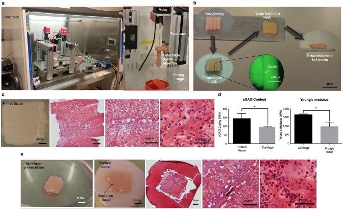
Figure 5. Bioprinting of cartilage tissue patches.
(a) Left: The homemade MABP in a flow cabinet. Right: A new print head design loaded with detachable nozzle assembly for tissue strand printing. (b) Images of printed tissue morphology over 3 weeks of incubation. (c) Characterization of a bioprinted cartilage 3 mm × 3 mm tissue patch showing Safranin-O/Fast Green at different magnifications. (d) sGAG content (n = 3) and Young’s modulus (for compression) (n = 4) of the bioprinted cartilage. (e) Implantation of 3D printed tissue patches into a 4 mm × 4 mm osteochondral defect with 2 mm thickness showing Safranin-O/Fast Green histology examination at different magnifications. All data are presented as average ± SD unless otherwise stated.

