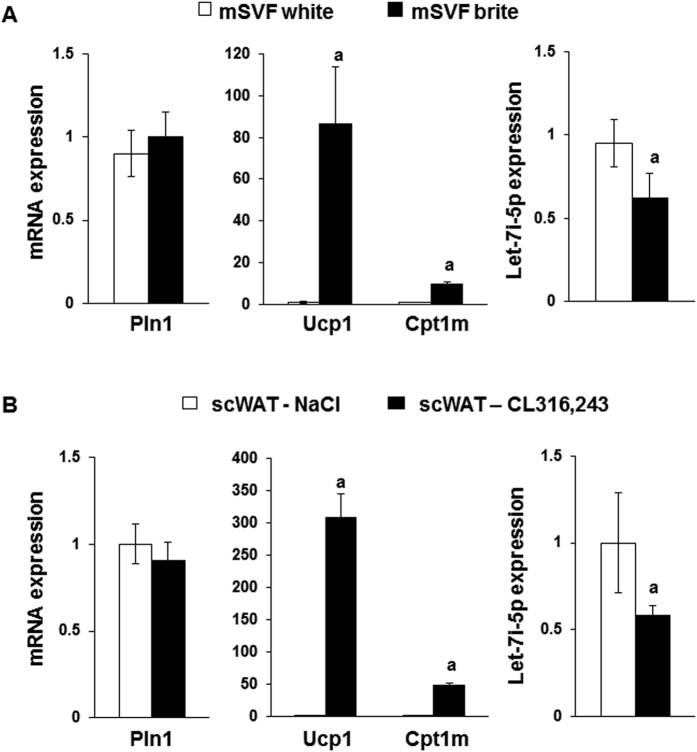
Figure 5. Let-7i-5p levels in mouse adipose tissue and cell models.
(A) Cells from SVF of scWAT were differentiated into white or brite adipocytes and used for mRNA and miRNA level quantification by RT-qPCR. (B) mRNA and miRNA expression determined by RT-qPCR in scWAT from C57BL/6 mice treated or not with CL316,243 for 1 week. Control animals were injected with vehicle (NaCl 0.9%, w/v). Histograms represent mean ± SEM of 3 independent experiments (A) of 8 mice (B). a: p < 0.05.

