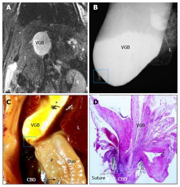
Figure 4.

Magnetic resonance imaging, microcholangiography and histomorphology of a rat on day 14 after surgically induced cholestasis towards creating a model of virtual gallbladder. A: Coronal T2-w MRI shows an oval-shaped hyperintense dilated CBD or VGB with L, K and S denoting the liver, kidney and stomach respectively; B: Digital microcholangiography displays a hyperdense dilated CBD or VGB with L denoting the liver; C: Laparotomic view shows the dilated CBD or VGB with L, P and Duo denoting the liver, pancreas and duodenum respectively; note the transparent distal CBD as indicated by a needle tip and an arrow, suggesting absent bile flow, and asterisk indicates where the needle hole was closed by suture ligation; D: Photomicrograph of hematoxylin and eosin stained slide of the ligature (dashed square on B and C) shows a complete CBD obstruction separating the VGB and distal CBD (original magnification × 100). MRI: Magnetic resonance imaging; CBD: Common bile duct; VGB: Virtual gallbladder.
