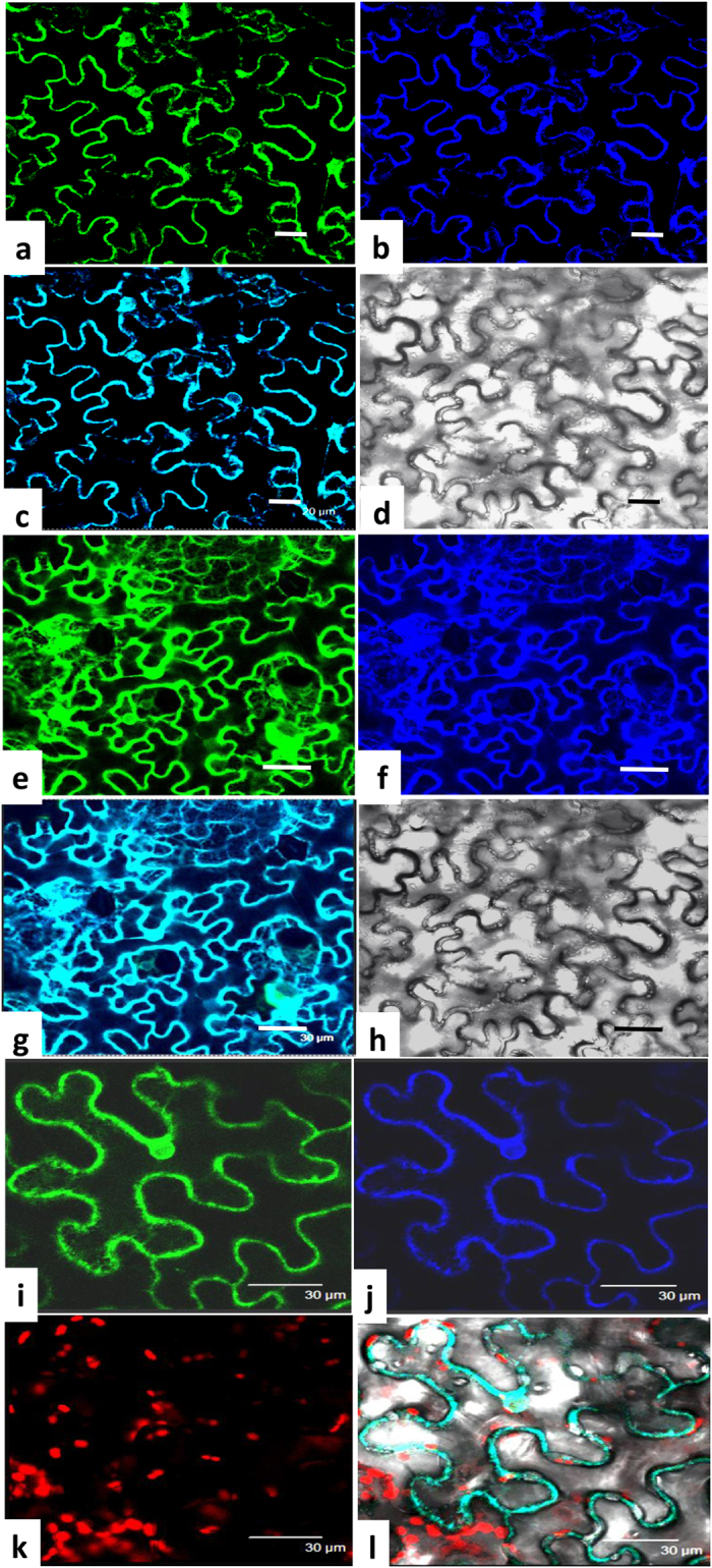
Figure 2. Subcellular localization of GmDGAT-1A and -2D.

GmDGAT1A-GFP, GmDGAT2D-GFP, and a ER marker CD3-959-mCherry, driven by the cauliflower mosaic virus 35S promoter, were transiently expressed in tobacco leaf epidermal cells. Materials were viewed by confocal microscopy. Represatative photos were shown. (a–d) Localization of GmDGAT1A-GFP. Image of GmDGAT1A-GFP (a), the blue fluorescence image of ER marker CD3-959-mCherry (b), and the merged image of GmDGAT1A-GFP and CD3-959-mCherry (c), and the bright field image (d). Bars = 20 μm. (e–h) Localization of GmDGAT2D-GFP. Image of GmDGAT2D-GFP (e), image of ER marker CD3-959-mCherry (f), the merged image of GmDGAT2D-GFP and ER marker CD3-959-mCherry (g), and the bright field image of cells (h). Bars = 30 μm. (i–l) Enlarged image of GmDGAT2D-GFP (i), image of ER marker (j), the chloroplast autofluorescence images (k), and merged image of GmDGAT2D-GFP, ER marker, and chloroplast autofluorescence in a bright field (l). Bars = 30 μm.
