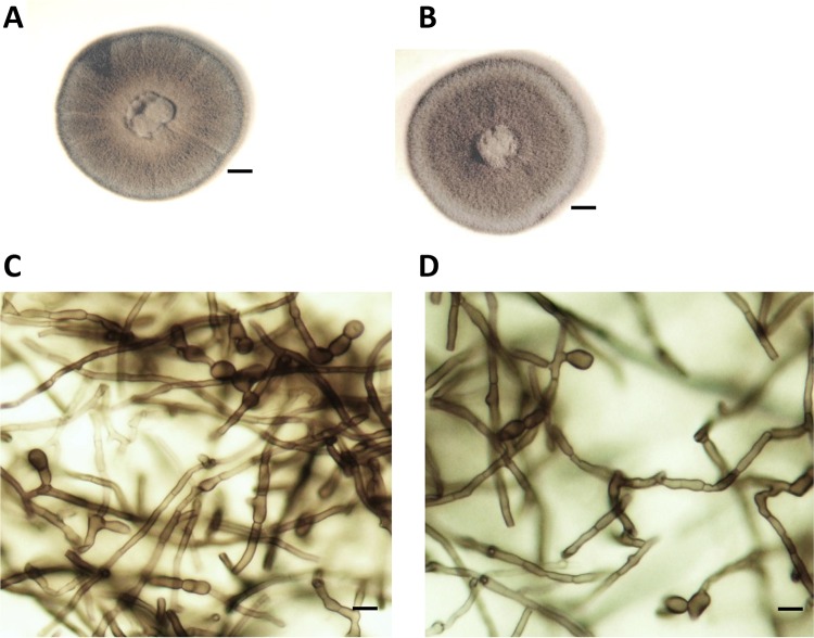
FIG 1.
Colonial and microscopic appearance of Emarellia grisea (NCPF 7066) (A and C) and E. paragrisea (NCPF 7611) (B and D). The colonial appearance after 2 weeks of culture on Sabouraud dextrose agar at 30°C (A and B) and the microscopic appearance showing lateral, terminal, and intercalary chlamydospores (prepared in lactophenol, ×400 magnification) are depicted (C and D). No discernible differences in colonial morphology or conidiation were noted on any media. Scale bar: 4 mm (A and C) or 5 μm (B and D).

