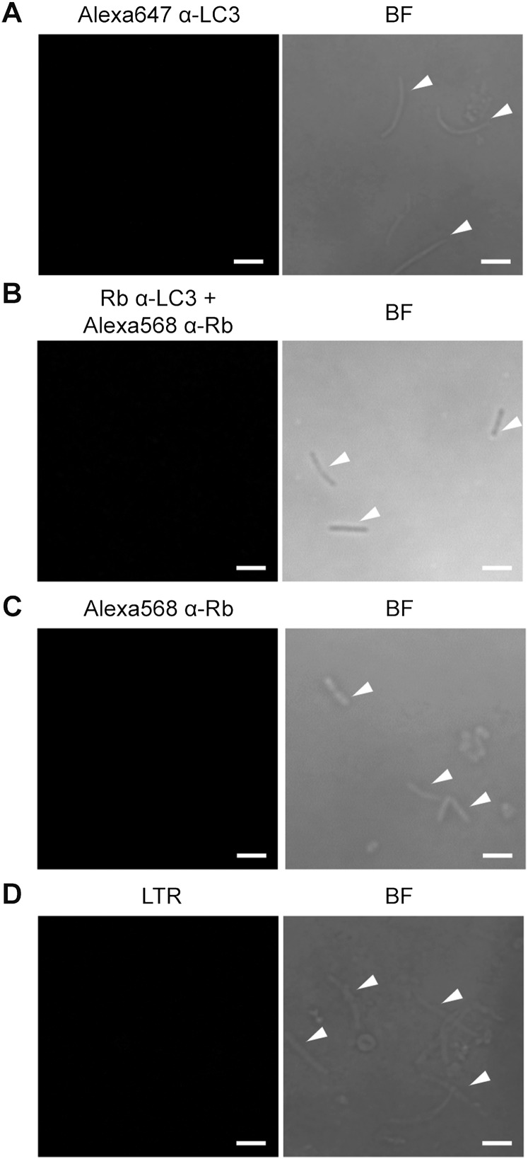
Fig. S4.
Anti-LC3 antibodies and LTR do not recognize isolated P. vivax sporozoites. (A–C) Isolated P. vivax sporozoites were spotted onto a 10-well antigen slide, fixed with 4% paraformaldehyde, and permeabilized with 0.1% saponin/3% BSA in PBS. The sporozoites were then stained with Alexa Fluor 647-conjugated rabbit anti-LC3 antibody at room temperature for 2 h (A). Alternatively, the sporozoites were stained with rabbit anti-LC3 antibody at 4 °C overnight, followed by Alexa Fluor 568-conjugated anti-rabbit secondary antibody at room temperature for 2 h (B) or with Alexa Fluor 568-conjugated anti-rabbit secondary antibody alone (C). The samples were then mounted and analyzed by confocal microscopy. (D) Isolated P. vivax sporozoites were incubated with LTR dye for 1 h at room temperature, washed twice with PBS, and spotted onto a 10-well antigen slide. The mounted samples were analyzed by confocal microscopy. Sporozoites are marked with white arrowheads. (Scale bar: 5 µm.)

