Fig. 2.
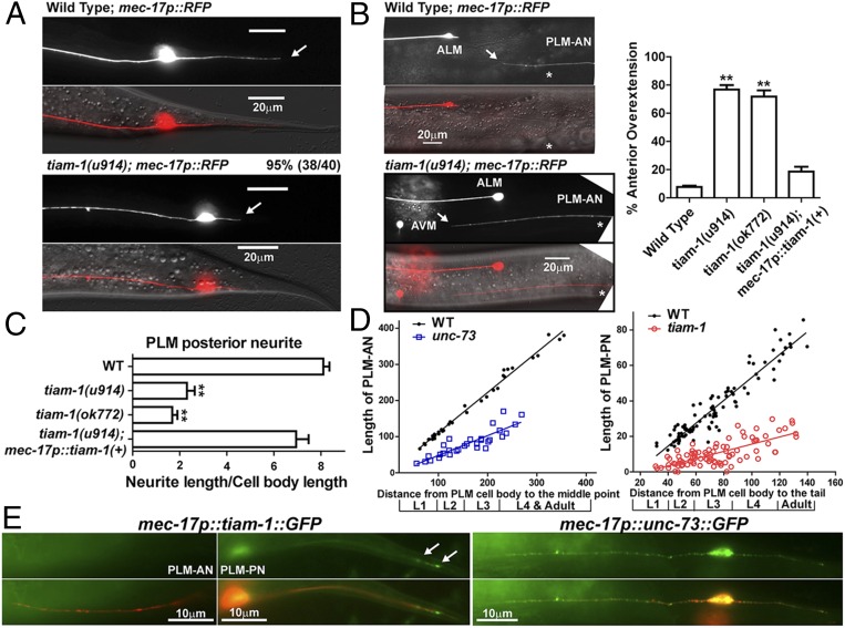
TIAM-1 promotes posteriorly directed neurite growth. (A) The shortening of PLM-PN in tiam-1 mutants compared with wild-type animals. Arrows indicate the end of PLM-PN. (B) Overextension of PLM-AN in tiam-1 animals and the percentage of PLM-AN passing the ALM cell body. Asterisks in the images indicate the position of the vulva, and the arrows point to where PLM-AN terminates. (C) The length of PLM-PN in tiam-1 animals. (D) The lengths (in μm) of PLM-AN and PLM-PN in wild-type, unc-73(u1031), and tiam-1(u914) animals during different developmental stages. The distance from the PLM cell body to the middle point (the position of vulva in L4 and adults) or the tip of the tail was used as a measurement of developmental progress. Linear regression (solid lines) was applied to these data. The slope indicates the growth rate of neurites relative to the extension of the worm body. For WT PLM-AN, the slope = 1.1 ± 0.02 and r2 = 0.99 (goodness of fit); for unc-73 PLM-AN, the slope = 0.55 ± 0.06 and r2 = 0.77; for WT PLM-PN, the slope = 0.65 ± 0.02 and r2 = 0.90; for tiam-1 PLM-PN, the slope = 0.2 ± 0.02 and r2 = 0.55. (E) The localization of TIAM-1::GFP and UNC-73::GFP fusion proteins expressed specifically in the TRNs from L1 animals that carry the mec-17p::RFP transgene. Arrows point to the position of TIAM-1::GFP in the distal region of PLM-PN.

