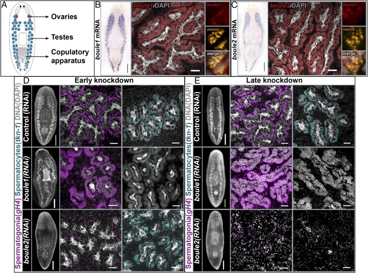
Fig. 1.
Planarian boule1 and boule2 perform different functions in spermatogenesis. (A) Illustration of sexual planarian depicting the positions of reproductive structures. Ovaries are in red, testes are in blue, and germ-line stem cells are in green. (B and C) Colorimetric ISH showing boule1 and boule2 mRNA expression in the testes. (Scale bars, 1 mm.) FISH detects boule1 and boule2 expression in spermatogonial stem cells (SSCs), spermatogonia, and spermatocytes. (Scale bars, 50 μm.) Coexpression of boule transcripts with nanos+ SSCs is shown. (D) Animals fixed following two feedings of dsRNA spaced 4–5 d apart. Control (RNAi), boule1(RNAi), and boule2(RNAi) animals labeled with germinal histone H4 (gH4) in magenta to detect mitotic spermatogonia and tektin-1 (tkn-1) in cyan to mark meiotic spermatocytes. boule1(RNAi) animals show absence of meiotic labeling, but expansion of spermatogonia. The spermatogonial layer is reduced in boule2(RNAi) animals, whereas the spermatocyte population is comparable to controls. (E) Animals fixed following four feedings of dsRNA spaced 4–5 d apart. boule1(RNAi) testes contain clusters of SSCs and spermatogonia; meiotic and postmeiotic male germ cells are absent. boule2(RNAi) animals show a loss of all male germ cells. The remaining gH4 label coincides with neoblasts (somatic stem cells). Left in D and E show whole-mount images. (Scale bars, 1 mm.) Middle and Right in D and E show high magnification view of testis lobes. (Scale bars, 50 μm.)

