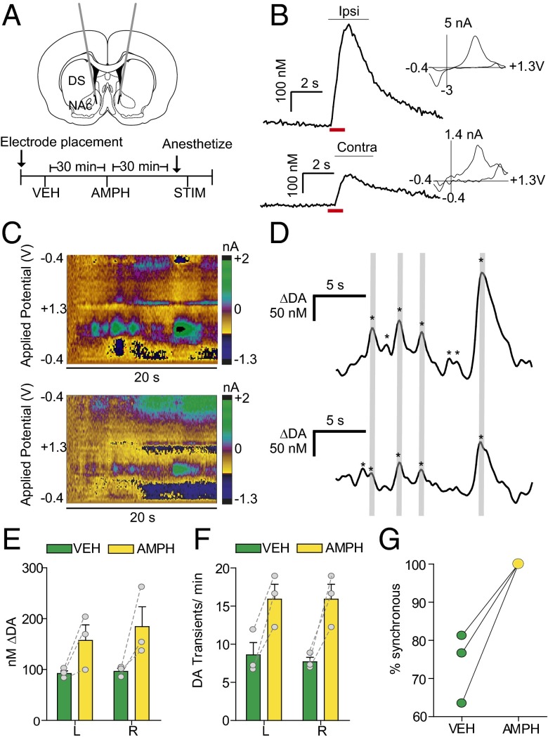
Fig. 1.
Spontaneous dopamine transients synchronize bilaterally in the NAc. (A) Schematic of electrode implantation for simultaneous dopamine transient measurements. (B) Representative dopamine release in the right and left NAc following stimulation (red bar) of the right VTA, with voltammograms. (C) Representative color plots demonstrating synchronous dopamine transients in both hemispheres with applied potential on the ordinate, recording time on the abscissa, and current encoded in false color. (D) Changes in dopamine concentration (asterisks) in both hemispheres extracted using principal component analysis from the color plots in C. Gray lines indicate synchronized transients; asterisks alone indicate asynchronous dopamine release. (E) Average ± SEM dopamine transient concentration in the left (L) and right (R) NAc after administration of saline vehicle (VEH; green) and 2.5 mg/kg amphetamine (AMPH; yellow). (F) Average ± SEM number of dopamine transients min−1 in the left and right NAc after VEH and AMPH administration. (G) Within-animal comparison of percent transient synchrony after VEH and AMPH administration. n = 3 animals.

