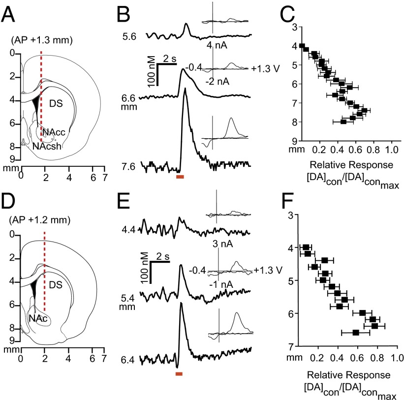
Fig. 2.
Stimulation of dopamine neurons produces localized release in the contralateral striatum. (A and D) Electrode trajectories superimposed on coronal sections. (B and E) Dopamine release at the terminals was dependent on recording electrode depth; example traces are shown. Cyclic voltammograms recorded at maximum release are shown as well. (C and F) Dopamine response (DAcon) following electrical stimulation of the contralateral VTA (C) or SN (F), normalized to maximum dopamine release (DAcon-max) as a function of working electrode depth. n = 10 animals per group. Data are average ± SEM.

