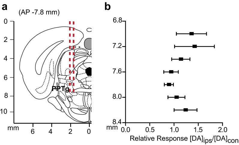
Fig. S1.
Stimulation of the PPTg elicits release in the contralateral DMS. (A) Coronal section showing stimulating electrode tracts in the PPTg. (B) Within-animal comparison of DMS effluxes resulting from ipsilateral stimulation (DAips) compared with contralateral stimulation (DAcon) at identical depths. n = 5 animals. Data are average ± SEM.

