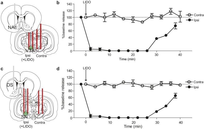
Fig. S3.
Contralateral dopamine release is not a result of electrical spread between hemispheres. (A and C) Schematics of carbon fiber electrodes in the NAc (A) and DMS (C), with a cannulated stimulating electrode in the ipsilateral VTA/SN for delivery of lidocaine and a stimulating electrode in the contralateral VTA/SN. (B and D) Dopamine release as a percentage of baseline in the NAc (B) and DMS (D), resulting from contralateral and ipsilateral stimulation of VTA/SN. Lidocaine (350 nmol/0.5 µL) delivery is indicated by the arrow. n = 4 animals per group. Data are average ± SEM.

