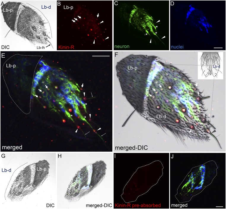
Fig. S8.
Confocal analyses of Aedae-KR immunolocalization in the distal segment of the labellum. In A, F, G, and H, DIC images show the labellum is composed of two segments, proximal and distal (dashed black lines in A, F, and G), where long labellar sensilla (long hair, black arrow in A) are present (see drawing in F). In C, E, F, H, and J neurons appear green (anti-HRP antibody). In D, E, F, H, and J nuclei appear blue (DAPI). The root fiber bundles in the ciliary region of sensory neurons are marked by asterisks in C and E. The receptor signal (red) is present in accessory cells at the base of long labellar sensilla, indicated by arrowheads in B, E, and F, and in accessory cells of short papilla, indicated by arrows in B, E, and F. No receptor signal was observed in the labellum proximal segment, the area enclosed by the dashed white line in E. Dendrites projecting toward the tip of the long labellar hair are shown in E and F (thin arrows). No receptor signal was observed in negative control tissues (antigen preabsorbed antibodies). G–J show the same tissue. Images as Z-stacks (Z-step: 0.66 µm) are as follows: A–F, 12 sections; G–J, 10 sections. Lb-d, distal segment of labellum; Lb-lh, long labellar hair; Lb-p, proximal segment of labellum. (Scale bars, 20 µm.)

