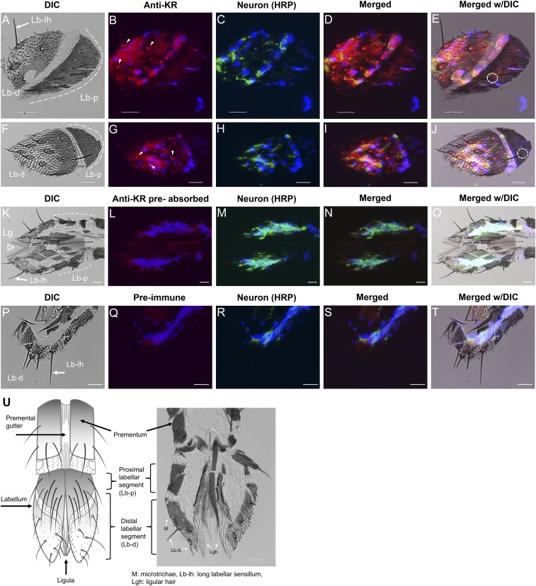
Fig. S9.
Immunofluorescence analyses of the Aedae-KR in female labellum. (A, F, K, and P) DIC images show the distal and proximal (dashed white lines) labellar segments and ligula (open arrowhead in K). (B and G) The receptor signal (red) was observed in sensory neurons of labellar sensilla (arrowheads) only in the distal labellar segment. (E and J) No receptor signal was observed in the proximal labellar segment, although hairs/sensilla were present (dashed circle). (L, N, O, Q, S, and T) No receptor signal was observed in tissues incubated with antigen preabsorbed antibody (L, N, and O) or with preimmune serum (Q, S, and T). In C, H, M, and R, neurons appear green. Merged images of receptor, neuron, and nuclear labeling (blue; DAPI) are shown in D, I, N, and S. Merged DIC images are shown in E, J, O, and T. (U) A schematic diagram of the labellum (28, 33) is compared with a labellar frozen section. Lb-d, distal segment of labellum; Lb-lh, long labellar hair; Lb-p, proximal segment of labellum; Lg, ligula; Lgh, ligular hair; M, microtrichae. (Scale bars, 20 µm.)

