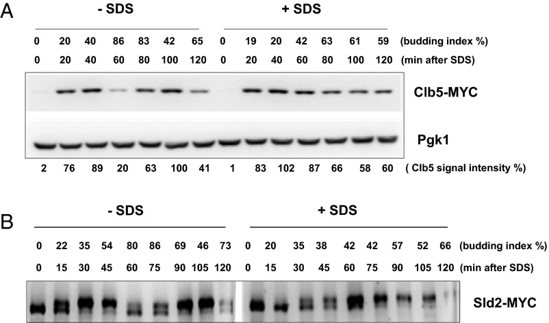
Fig. S1.
Clb5 is stabilized after SDS treatment. (A) CLB5-MYC cells were synchronized during G1 phase by α-factor. The cell cycle block was released and SDS was added to the media. Samples were collected every 20 min for protein extractions. Each time point was subjected to Western blot analysis. Pgk1 was used as a loading control. Budding index is shown as percent of budded cells. (B) SLD2-MYC cells were used to monitor the Sld2 phosphorylation status upon G1 block and release as described in A.

