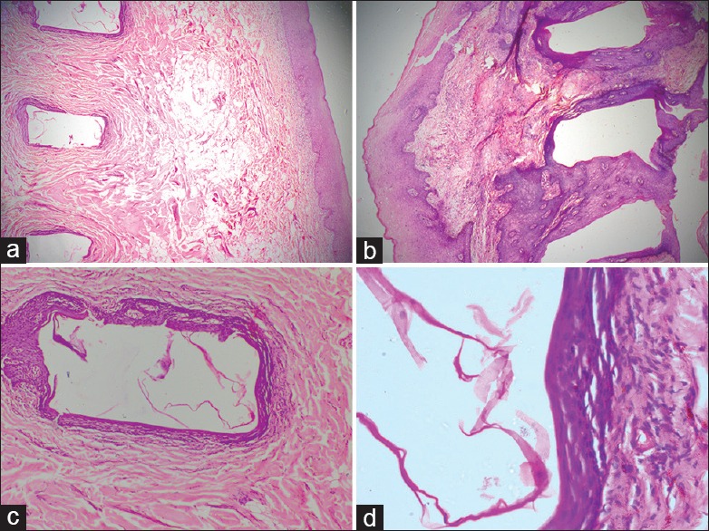
Figure 1.

(a and b) Both right and left side linearly arranged multiple linearly arranged epidermoid cysts with overlying stratified squamous epithelial clusters (H and E, ×40); (c) Section shows cyst lined by stratified squamous epithelium with granular cell layer. No appendages are noted within the wall of cyst (H and E, ×100); (d) Cyst wall lined by orthokeratotic stratified squamous epithelium with keratin flakes in the lumen (H and E, ×400)
