
Fig. 3.
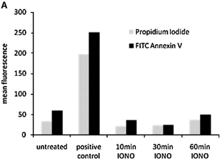

Ionomycin leads to ErbB1 trafficking to MVB compartments. A: HaCaT were treated with 1 mM ionomycin for the indicated length of time and cells were analyzed for apoptosis using Annexin-V and PI staining and FACS analysis. Treatment with staurosporine (1 μM) for 16 h served as positive control. B, C: HaCaT cells were treated with ionomycin (1 μM) for 10, 30, and 60 min and cell homogenates layered on a step sucrose gradient as described above. Homogenate gradient fractions and exosomes isolated from CM were analyzed by Western blot using the anti-ErbB1 C-terminus antibody. D: HaCaT cells were grown on glass cover slips and treated with or without ionomycin (1 μM) for 20 min. Cells were fixed and permeabilized in 3% paraformaldehyde/Triton X100 and stained with a mouse anti-ErbB1 ectodomain antibody followed by a Cy3-conjugated antimouse IgG secondary antibody. Confocal images were taken in the DKFZ microscopy division. [Color figure can be viewed in the online issue, which is available at www.interscience.wiley.com.]