
Figure 5. HNF4γ and NR2F2 protein expression in gastric cancer cell lines.
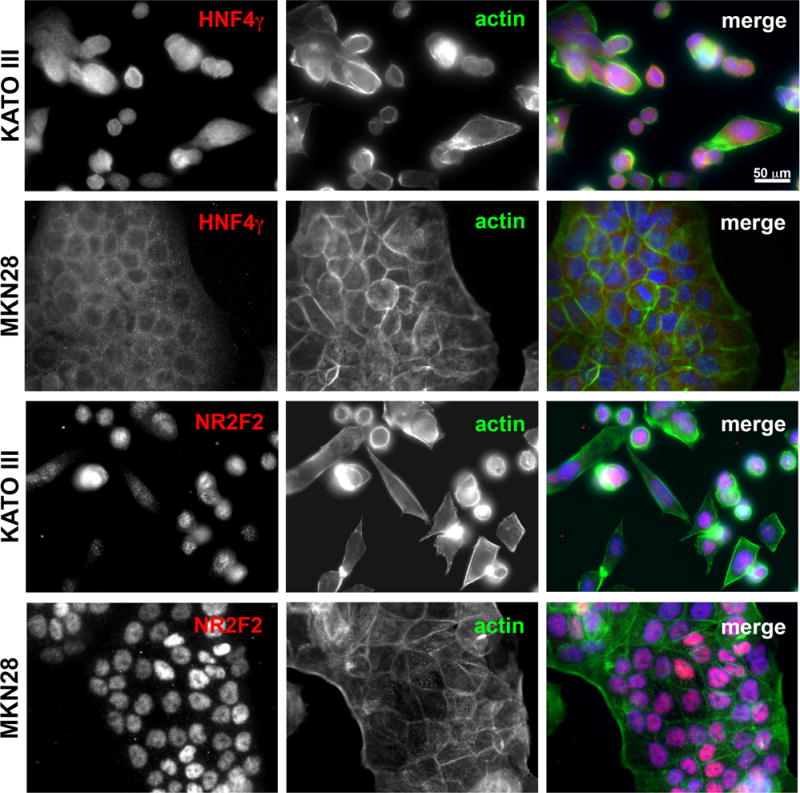
Immunofluorescence using HNF4γ or NR2F2 specific antibodies (red) in the gastric cell lines KATO III and MKN28. A co-labeling with Alexa488-conjugated phaloidin and DAPI was performed for better visualization of cell and nuclear morphology. Images were taken under 40X objective in a Zeiss Axiophot microscope.