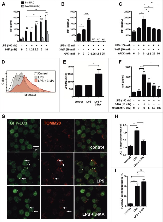
Figure 2.

MIF secretion in autophagy-deficient cells is dependent on mitochondrial reactive oxygen species (ROS). (A) PMA-differentiated THP-1 cells were treated with LPS (100 ng/ml) and different concentrations of 3-methyladenine (3-MA) in the presence or absence of the ROS inhibitor N-acetyl-L-cysteine (NAC, 25 mM) for 6 h. Secreted MIF was measured by ELISA. (B and C) Immortalized bone marrow-derived macrophages (iBMM) were treated with LPS (100 ng/ml) and 3-MA (10 mM) in the presence or absence of different concentrations of (B) NAC or (C) the ROS inhibitor (2R,4R)-APDC (APDC). Bars represent means ± SEM of triplicates, representative of at least 3 experiments. (D) RAW264.7 cells were treated with LPS (100 ng/ml) or LPS + 3-MA (10 mM) for 6 h, then incubated with MitoSOX Red to measure mitochondrial superoxide production by FACS. The geometric mean fluorescence index (MFI) is shown in (D and E). (F) iBMM were treated with LPS + 3-MA ± MitoTEMPO for 6 h at the concentrations stated and MIF secretion measured by ELISA. (G) iBMM stably transfected with GFP-LC3 were treated with LPS (100 ng/ml) or LPS + 3-MA (10 mM) for 6 h, fixed and stained with antibody against TOMM20. Autophagosome formation (H) and percentage of TOMM20+ autophagosomes (I) were analyzed. Bars represent means ± SEM of 3 separate experiments. *, p < 0.05; **, p < 0.01; ***, p < 0.005; ****, p < 0.001.
