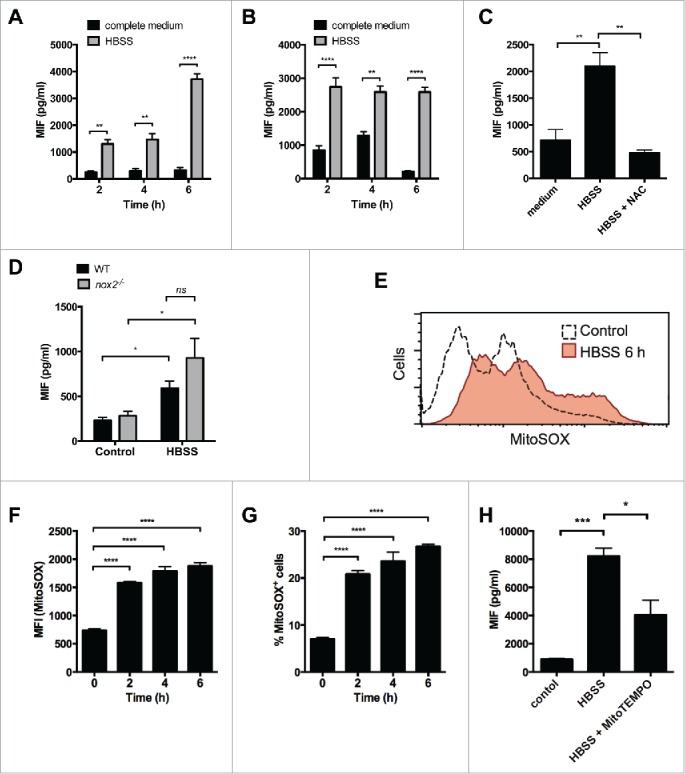
Figure 3.

Amino acid starvation induces MIF secretion by macrophages. (A) PMA-differentiated THP-1 cells or (B) RAW264.7 cells were starved in Hank's balanced salt solution for the times stated and secreted MIF measured by ELISA. (C) PMA-differentiated THP-1 cells were starved in Hank's balanced salt solution (HBSS) for 6 h in the presence or absence of the ROS inhibitor N-acetyl-L-cysteine (NAC, 25 mM) and secreted MIF measured by ELISA. (D) Primary BMM from WT and cybb−/− mice (n = 3) were treated with HBSS for 6 h and MIF secretion measured by ELISA. (E) FACS analysis (geometric mean) of MitoSOX in RAW264.7 cells treated with HBSS for 6 h. Geometric mean fluorescence for MitoSOX (F) and percentage of MitoSOX+ RAW264.7 cells (G) after treatment with HBSS over time. (H) iBMM were treated with HBSS or HBSS + MitoTEMPO (5 nM) for 6 h. MIF secretion was measured by ELISA. Bars represent means ± SEM of 3 separate experiments. *, p < 0.05; **, p < 0.01; ***, p < 0.005; ****, p < 0.001.
