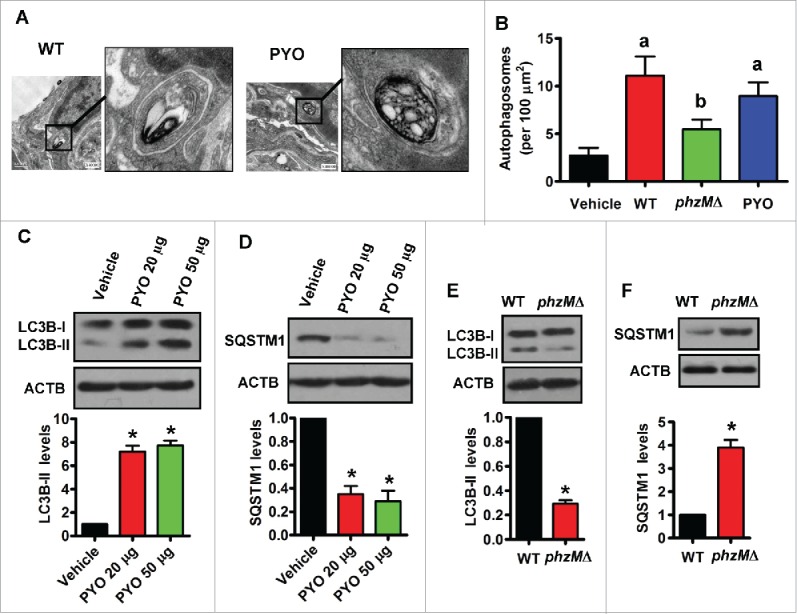
Figure 3.

Pyocyanin induces autophagy in lung tissues. (A) After rats were inoculated intratracheally with wild-type (WT) P. aeruginosa or pyocyanin (50 μg), the lung tissues were collected and fixed, and processed for transmission electron microscopy study. Representative images of the lung tissues from rats treated with WT P. aeruginosa (left) and pyocyanin (PYO) (right) are shown. (B) The numbers of autophagosomes were counted. Data are expressed as mean ± SD of 3 independent experiments. a, P < 0.05 vs. vehicle; b, P < 0.05 vs. WT. (C and D) After rats were inoculated intratracheally with pyocyanin, the protein levels of LC3B (C) and SQSTM1 (D) were determined in lung tissues by western blotting. The blot is representative of 3 experiments. The lower panels show quantification of the ratio of LC3B-II or SQSTM1 to ACTB. *, P < 0.05 vs. vehicle. (E and F) After rats were inoculated intratracheally with P. aeruginosa, the protein levels of LC3B (E) and SQSTM1 (F) were determined in lung tissues. The blot is representative of 3 experiments. The lower panels show quantification of the ratio of LC3B-II or SQSTM1 to ACTB. *, P< 0.05 vs. WT. Bars, 0.5 μm.
