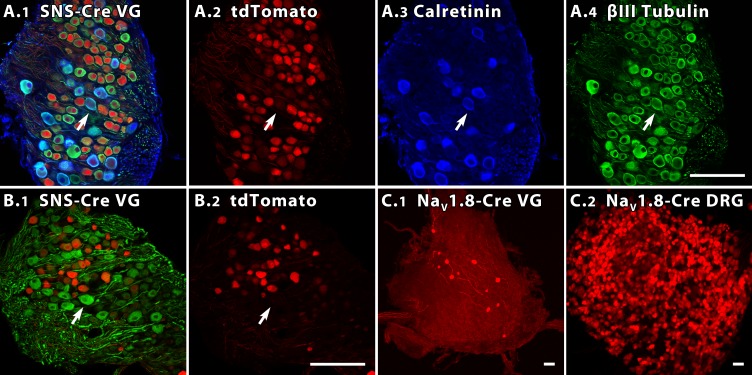
Fig. 10.
In 2 NaV1.8 reporter mouse lines, different percentages of vestibular ganglion neurons are labeled. A and B: 35-μm sections from the superior vestibular ganglia (VG) of 4- to 6-wk SNS-Cre reporter mice, showing tdTomato (red) in many VGNs. Scale bars (50 μm) in A4 and B2 apply to A1-A4 and B1-B2. A1: SNS-Cre VG colabeled with antibodies to calretinin (blue) and β-III tubulin (green). A2-A4: color channels of A1 shown separately. Calretinin-positive cells (A3) are relatively large and not tdTomato-positive (e.g., arrow). B1: SNS-Cre section from a different mouse, colabeled with NF200 antibody (green). Intensely NF200-positive cells (e.g., arrow) are often large and not tdTomato-positive. B2: same section showing tdTomato alone. C: tdTomato expression in VG (C1) and DRG (C2) of a 5- to 6-wk NaV1.8-Cre reporter mouse. Scale bars = 50 μm. C1: maximum intensity projection image of the VG. C2: whole mount DRG from the same animal. Despite strong tdTomato label in the DRG, fewer VGNs are positive in this reporter line than in the SNS-Cre mouse.

