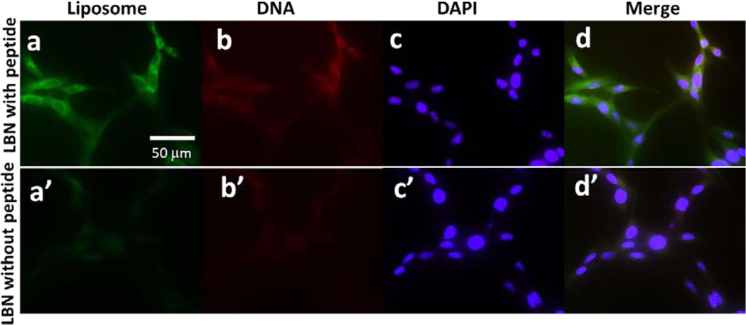
Figure 3.
Fluorescent images of rMSCs after internalization of LBNs with and without 3VT-peptide for 4 h. Lipids, DNA, and cell nuclei were labeled with a green dye (carboxylfluorescein, a and a′), a red dye (rhodamine, b and b′), and DAPI (c and d′), respectively; d is a merged version of a, b, and c; d′ is a merged version of a′, b′, and c′.

