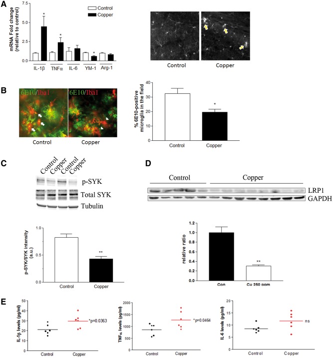
FIG. 7.
Aberrant activation of neuroinflammation in the brain of copper-exposed 3xTg-AD mice. Three months old 3xTg-AD mice were treated with 250 ppm copper-containing drinking water for 3–12 months. A, The mRNA expression of selected cytokines and phagocytosis markers in the brain after the 3 months of control water (n = 10, open bars) or copper exposure (n = 10, closed bars). *P < 0.05 compared to the control group. Representative immunofluorescent staining of Iba1-positive microglia in hippocampus of 3xTg-AD mice received control or copper-containing drinking water for 3 months shows early signs of microglial activation. Arrows indicate the clustering and activating microglia in the absence of Aβ plaques in the copper-exposed mice. B, Representative double immunofluorescent staining of Iba1-positive microglia (red) and 6E10-positive Aβ plaques (green) in hippocampus of 3xTg-AD mice received control or copper-containing drinking water for 9 months. Arrows indicate microglia colocalized with 6E10-positive Aβ, and arrowheads indicate microglia located nearby Aβ plaques but no obvious 6E10-positive Aβ within. The graph represents the percentage of Iba1-positive microglia that colocalize 6E10 positive Aβ. *P < 0.05 compared to the control group (n = 5–8). C, The activation of SYK was determined by immunoblot of phospho-SYK and total SYK in the brain homogenates from 9 months treatment (**P < 0.01 compared to control, n = 4). D, The down-regulation of brain LRP1 was detected in 3xTg-AD mice exposed to copper-containing drinking water for 12 months (**P < 0.01, control n = 5, copper-exposed n = 8). E, Brain cytokine levels were measured by ELISA following 12 months copper exposure (n = 6).

