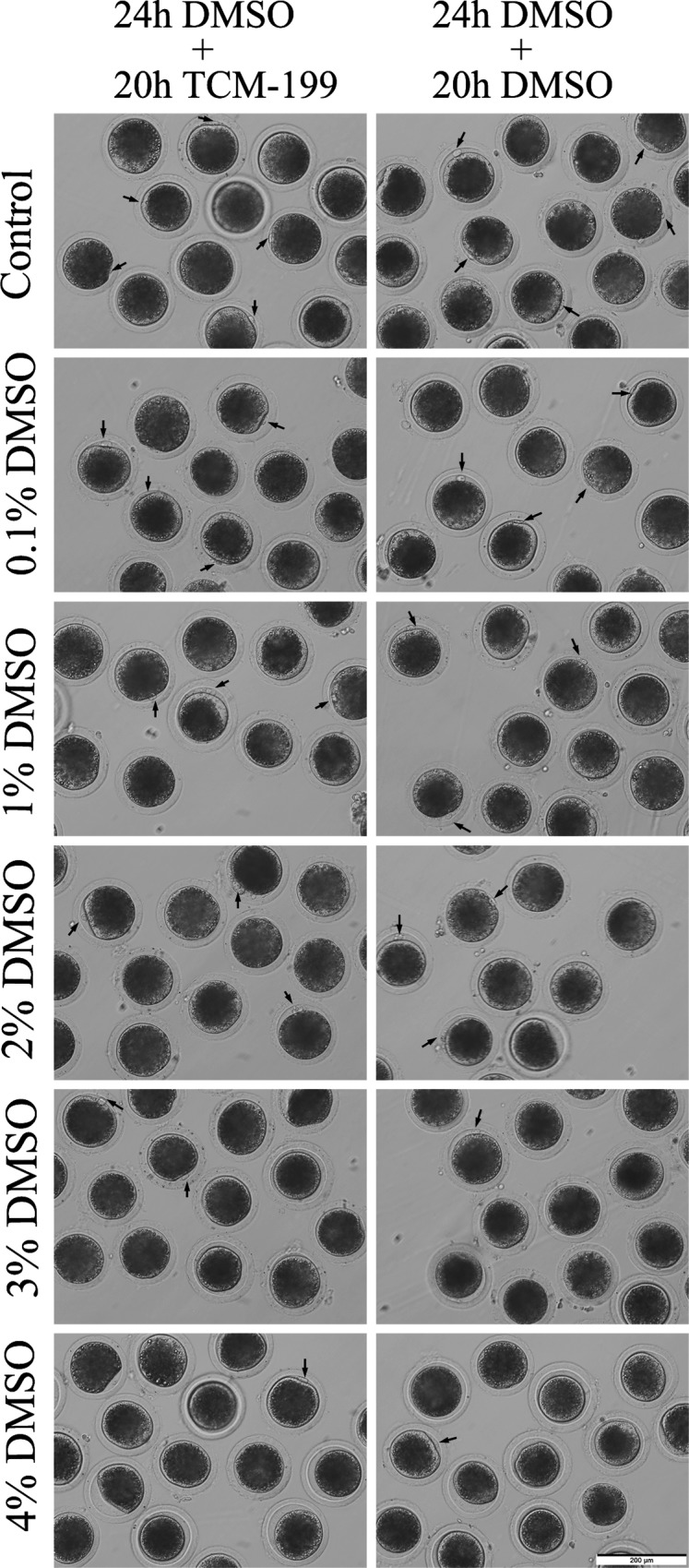
Fig 3. Morphology of oocytes after DMSO treatment.

COCs were cultured in vitro with maturation medium supplemented with 0%, 0.1%, 1%, 2%, 3% and 4% DMSO for 24h, and then transferred into without (termed as the 24h DMSO+20h TCM-199 group) or with DMSO (termed as the 24h DMSO+20h DMSO group) maturation medium to mature for 20h. Then cumulus cells were stripped off to take images for denuded oocytes. Arrows indicated the normal sized first polar bodies of MII oocytes. Scale bar, 200μm.
