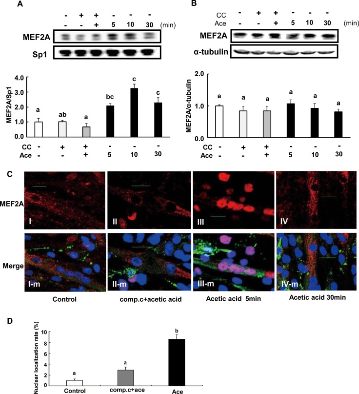
Fig 7. Effect of acetic acid treatment on nuclear MEF2A expression in L6 myotube cells.
L6 myotube cells were treated with 0.5 mM acetic acid, and nuclear fraction (A) and cytosolic fraction (B) were separated. MEF2A level was examined by western blotting as described in materials and methods. L6 myotube cells were cultured on glass cover slips coated with poly-L-lysine and treated with 0.5 mM acetic acid in the presence or absence of 10 μM compound C (C). Then cells were fixed and nuclear DNA was stained by Hoechst 33258 (blue). Cells were immunostained for MEF2A (red) and myosin (green). Scale bar = 20 μm. The nucleus immunostained with anti-MEF2A antibody were counted (8 mm2 area, n = 3) and the rate of nuclear localization of MEF2A was calculated (D). Each bar represents the mean ±SE (n = 3–4). Results were analyzed with one-way ANOVA followed by the Tukey-Kramer post hoc test for multiple comparisons. Groups without the same letter are significantly different (p<0.05).

