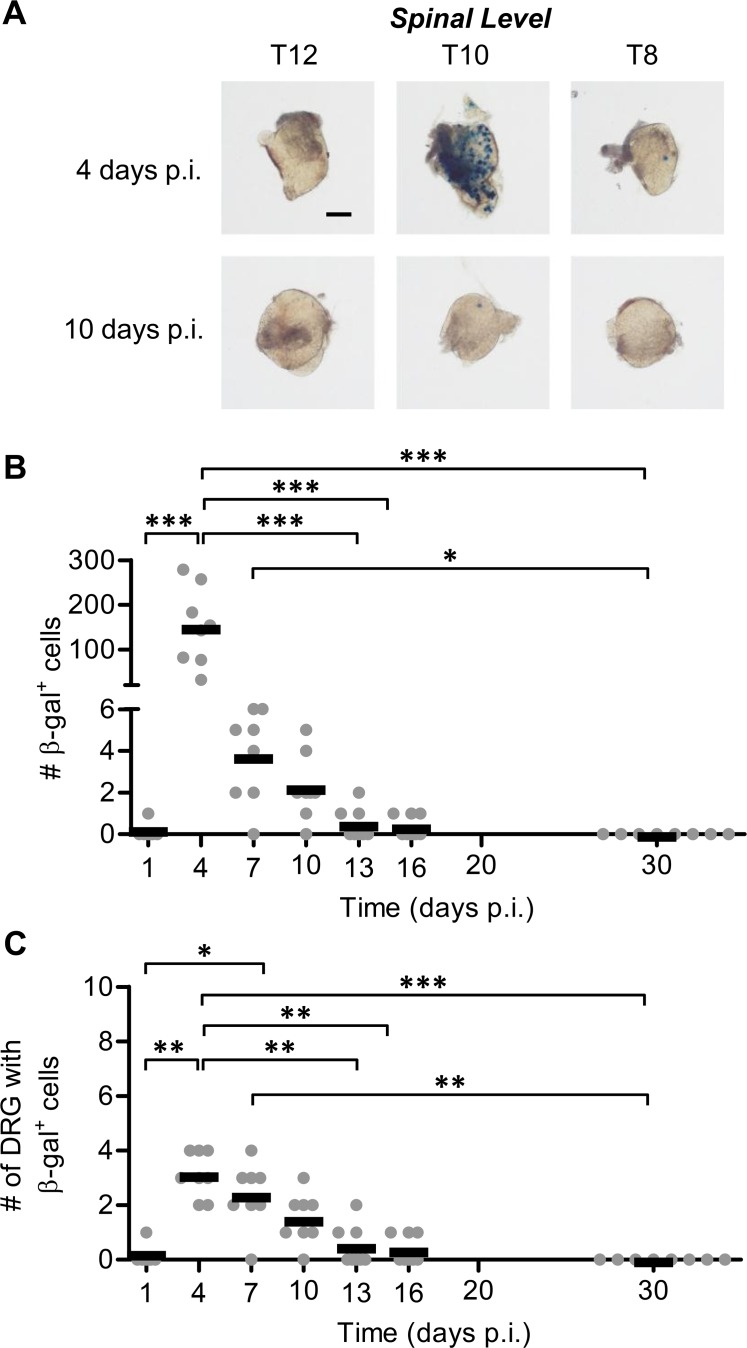
Fig 3. Conventional reporter genes show only minor lytic gene expression after the peak of acute infection and none in latency.
Groups of C57Bl/6 mice were infected with KOS6.β by tattoo on the flank and were culled at the indicated times p.i. for the determination of the number of β-gal+ cells per DRG. (A) Representative photomicrographs of DRG at spinal levels T12, T10 and T8 of a single mouse for either 4 or 10 days p.i. taken at 40× magnification (scale bar = 300 μm, as indicated on the top left image). (B) The total number of β-gal+ cells per mouse was estimated. (C) The spread of virus as determined by the number of DRG which contain at least one β-gal+ cell. The results of two independent experiments are pooled (n = 8), with each point representing a single mouse and the bar representing the mean cell count. (**p < 0.01, ***p < 0.001).

