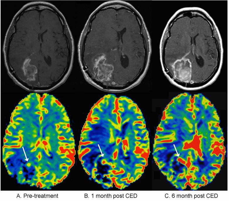
Figure 3.
Patient with true progressive disease, meaning an increase in contrast-enhancing tumor volume was observed within the first five months after treatment, and the patient had progressive disease at six months. A) Pretreatment contrast enhanced T1 weighted sequence (top) and corresponding cerebral blood volume map (bottom). The rCBV map color scale is weighted with red representing the highest values and black the lowest values. The white arrow indicates the portion of enhancing tumor with the highest rCBV. B) one-month post-treatment: an increase in eTV is noted with a corresponding increase in rCBV C) six months post treatment, showing progressively increasing eTV compatible with progressive disease as well as increasing rCBV.

