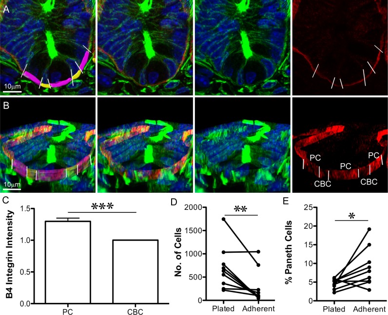
Fig 4. Paneth cells are more adherent to laminin than other crypt cells.
(A, B) Immunofluorescence images of sectioned tissue show nuclei (Hoechst, blue), F-actin (Phalloidin, green), and β4 Integrin (red): (A) is an optical section; (B) is a 3D projection (from Imaris) of the same crypt. In the left panel, the basal surface of crypt base columnar (CBC) cells are pseudo-coloured yellow, and the basal surface of Paneth cells are pseudo-coloured purple. Paneth cells are identifiable as the large cells with round, basally placed nuclei; CBC cells are narrow and have compressed nuclei. White lines (left and right panels) indicate the boundaries between Paneth cells and their neighbours. (C) Average β4 Integrin intensity on the basal surface of Paneth cells (PC) was normalised to the average value for neighbouring cells (CBC). Left panels in (A) and (B) indicate the Imaris-rendered surfaces that were used to measure β4 Integrin signal intensity. ± SEM, p < 0.001 (t test), n = 59 Paneth cells (S2 Data.) (D) The number of mouse small intestinal crypt cells attached to laminin-coated surfaces before (Plated) and after (Adherent) shaking decreases (p = 0.0078, Wilcoxon’s paired t test of eight independent experiments, S3 Data). (E) The proportion of attached cells from (D) staining positive for Lysozyme increases after shaking (p 0.0195, Wilcoxon’s paired t test of eight independent experiments, S3 Data). Underlying data for panel C can be found in S2 Data, and for panels D and E in S3 Data.

