
Fig. 3.
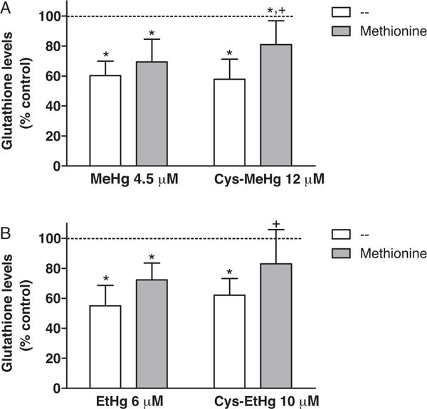
Glutathione levels after mercurial exposure. Rat C6 glioma cells were treated with MeHg, MeHg-Cys (A), EtHg, or EtHg-Cys (B) in HBSS (at concentrations approximating their respective EC50 value in the toxicity assay – Fig. 1). L-Methionine was added to the incubation medium (HBSS) 15 min before the mercurial’s addition at a final concentration 1000 fold greater than the Hg-compounds. After 30 min, HBSS was replaced by fresh DMEM and the cells were incubated for additional 4 h at 37 °C in a humidified atmosphere of 5% CO2. GSH concentration in control C6 cells (non treated) was 69.0 ± 2.2 nmol of GSH/mg of protein. GSH levels are expressed as percentage of control values (100%, dashed line). The asterisks (*) mean significant differences (p < 0.01) when compared to control and the symbol + means significant difference (p < 0.05) when compared to cells treated only with the respective mercurial (without Cys) by two-way ANOVA followed by the Bonferroni test. Data are expressed as mean ± SEM (N = 4 independent experiments).