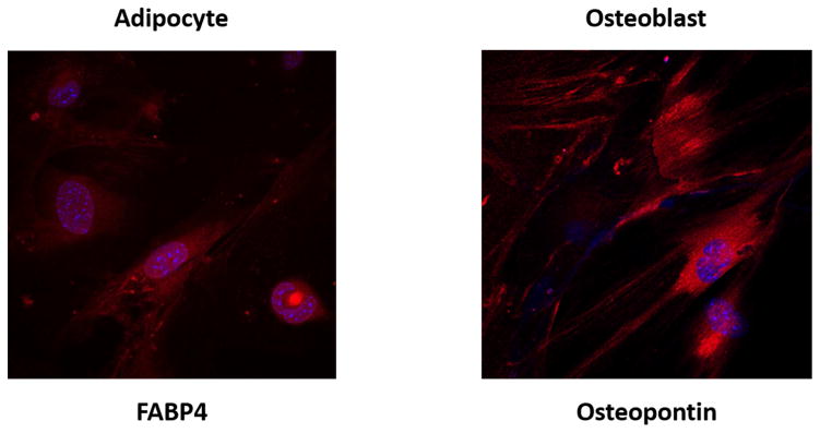
Figure 2. Adipose-derived mesenchymal stem cells (ASCs) isolated from 4 month and 22 month old male C57BL/6 mice demonstrate adipogenic and osteogenic differentiation potential.
The mouse mesenchymal stem cell functional identification kit (R&D Systems, Minneapolis, MN) was used to stimulate differentiation of ASCs into adipocytes (left) and osteoblasts (right), per the manufacturer’s instructions. Briefly, adipocytes were stained using goat anti-mouse FABP4 primary antibody, and osteocytes were stained using goat anti-mouse osteopontin primary antibody. Subsequently, both were exposed to donkey anti-goat IgG affinity purified PAb NorthernLights 557 fluorochrome-labeled secondary antibody (R&D Systems). Immunocytochemical staining of differentiated cells are presented with DAPI (blue) and NorthernLights (red) fluorescence imaging at magnification 40x.

