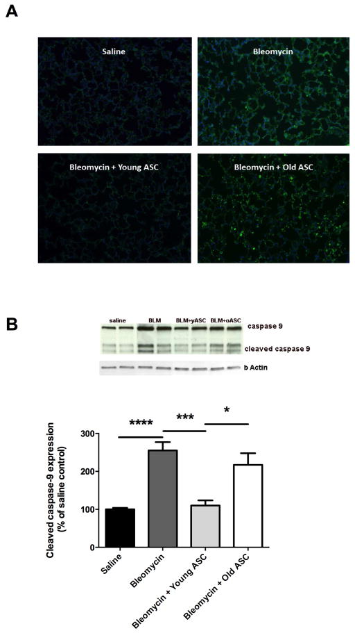
Figure 6. Markers of apoptosis are increased in lung tissue of aged male C57BL/6 after bleomycin (BLM) fibrosis, and inhibited by infusion of young adipose derived mesenchymal stem cells (yASC) but not old ASCs (oASC).
A) ApopTag assay was performed on lung tissue sections from aged male C57BL/6 mice receiving saline, bleomycin (BLM), BLM+yASC, or BLM+oASC. Panels are stained with FITC (nuclear TUNEL labeling) and DAPI (nuclear staining) as indicated in the Methods section. B) Western blots were performed on lung tissue protein extracts from aged male C57BL/6 mice receiving saline, BLM, BLM+yASC, or BLM+oASC to measure the ratio of cleaved caspase-9 relative to total caspase-9. A representative Western blot is shown of two individual mice/group. Data are graphed as the mean ± SEM of n=4–10/group.* p<0.05, ***p<0.001, ****p<0.0001.

