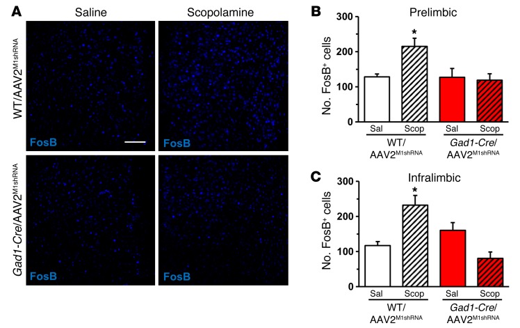
Figure 4. M1-AChR knockdown in Gad1-Cre mice decreased FosB activation following scopolamine treatment.
WT or Gad1-Cre mice received bilateral infusion of AAV2M1shRNA in the mPFC. Following behavioral tests, mice received an acute scopolamine injection (25 μg/kg) and were perfused 1 hour later. Brains were collected and processed for immunohistology. (A) Representative images of FosB labeling in the prelimbic mPFC of WT/AAV2M1shRNA and Gad1-Cre/AAV2M1shRNA mice treated with saline or scopolamine. Scale bar: 100 μm. (B) Quantification of FosB+ neurons in the (B) prelimbic or (C) infralimbic mPFC. Bars represent the mean ± SEM, n = 4–5/group. *P < 0.05, means significantly different from respective saline group based on ANOVA.

