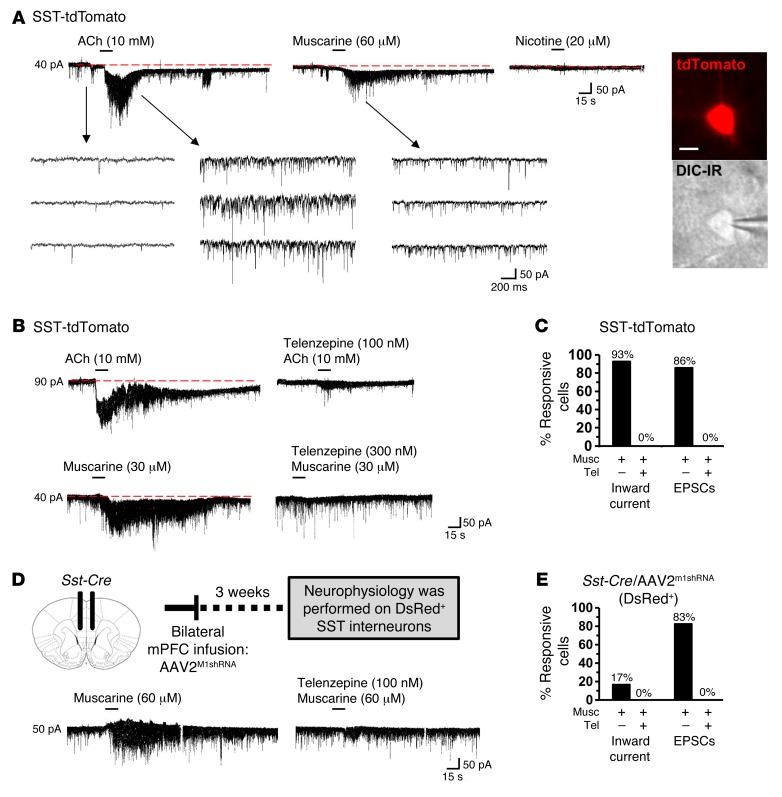
Figure 6. M1-AChR mediates cholinergic stimulation of SST interneurons in the mPFC.
SST-tdTomato and Sst-Cre/AAV2M1shRNA mice were used for brain slice electrophysiology. (A) Representative electrophysiology traces from SST-tdTomato interneurons in the mPFC following application of ACh, muscarine, or nicotine. Consecutive traces shown in the lower panel depict the spontaneously occurring excitatory, inward synaptic currents recorded at –70 mV in the absence and presence of agonists. Representative image of SST-tdTomato neuron with whole cell patch clamp. Scale bar: 10 μm. (B) Representative electrophysiology traces from SST-tdTomato interneurons in the mPFC after application of ACh alone or telenzepine followed by ACh. Lower traces show SST-tdTomato interneurons in the mPFC after application of muscarine alone or telenzepine followed by muscarine. (C) Proportion of SST-tdTomato interneurons that exhibited inward current and EPSCs following stimulation with muscarine (n = 14) or telenzepine followed by muscarine (n = 6). (D) Schematic and representative electrophysiology traces from DsRed+ interneurons in the mPFC of Sst-Cre/AAV2M1shRNA mice following application of muscarine alone or telenzepine followed by muscarine. (E) Proportion of DsRed+ interneurons that exhibited inward current and EPSCs after stimulation with muscarine (n = 6) or telenzepine followed by muscarine (n = 3).

