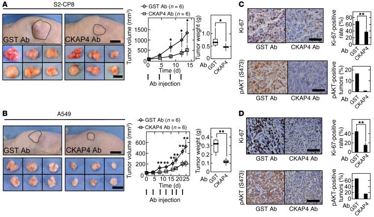
Figure 10. Antiproliferative effect of anti-CKAP4 antibody on DKK1- and CKAP4-expressing cancer cells in vivo.
(A and B) S2-CP8 cells (A) and A549 cells (B) were implanted s.c. into immunodeficient mice. Anti-CKAP4 (150 μg/body) (n = 6) or GST antibody (150 μg/body) (n = 6) was injected into the intraperitoneal cavity twice per week. Left panels: Representative appearance of 1 mouse (top picture) and extirpated xenograft tumors (bottom picture) are shown. The volume (middle panel) and weight (right panel) of the xenograft tumors were measured. (C and D) Sections prepared from xenograft tumors of S2-CP8 cells (C) and A549 cells (D) were stained with hematoxylin and anti–Ki-67 (top panel) or anti-pAKT (bottom panel) antibody. Ki-67–positive cells were counted, and the number is expressed as the percentage of total cells per field (n = 5 fields) in the right panel. Percentages of pAKT (S473)–positive tumors in the total xenograft tumors tested are shown in the right panel. Results are shown as means ± SD of 3 independent experiments. *P < 0.05; **P < 0.01 (2-tailed Student’s t test). Scale bars: 10 mm (A and B); 100 μm (C and D).

