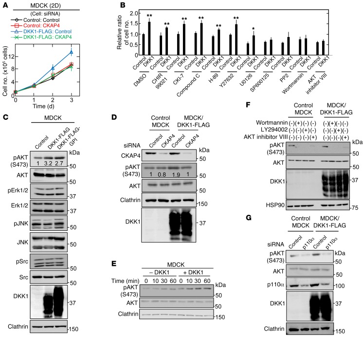
Figure 3. DKK1 signaling through CKAP4 activates the PI3K/AKT pathway, resulting in cellular proliferation.
(A) Control MDCK and MDCK/DKK1-FLAG cells were transfected with siRNA for control or CKAP4, and the cells were then subjected to the 2D cell proliferation assay. Results are shown as the mean ± SD of 3 independent experiments. (B) Control MDCK and MDCK/DKK1-FLAG cells were cultured at densities of 1 × 10⁵ cells in a 35-mm dish 2-dimensionally for 60 hours. The cells were treated for the last 36 hours with 10 μM CHIR99021, 100 μM CKI-7, 10 μM Compound C, 10 μM H-89, 1 μM Y27632, 10 μM U0126, 10 μM SP600125, 10 μM PP2, 200 nM Wortmannin, or 5 μM AKT inhibitor VIII, and then cell numbers were enumerated. Relative cell numbers are shown as fold changes compared with those in DMSO-treated control MDCK cells. Results are shown as the mean ± SD of 3 independent experiments. *P < 0.05; **P < 0.01 (2-tailed Student’s t test). (C) Lysates of control MDCK, MDCK/DKK1-FLAG, or MDCK/DKK1-GPI-FLAG were probed with the indicated antibodies. Clathrin was used as a loading control. (D) Control MDCK or MDCK/DKK1-FLAG cells were transfected with control or CKAP4 siRNA, and cell lysates were probed with the indicated antibodies. Clathrin was used as a loading control. (E) MDCK cells were stimulated with 10 nM DKK1 for the indicated time periods, and cell lysates were probed with the indicated antibodies. (F) Control MDCK or MDCK/DKK1-FLAG cells were treated with 200 nM Wortmannin, 50 μM LY294002, or 5 μM AKT inhibitor VIII for 30 minutes, and cell lysates were probed with the indicated antibodies. (G) Control MDCK or MDCK/DKK1-FLAG cells were transfected with control (scramble) or p110α siRNA, and cell lysates were probed with the indicated antibodies.

