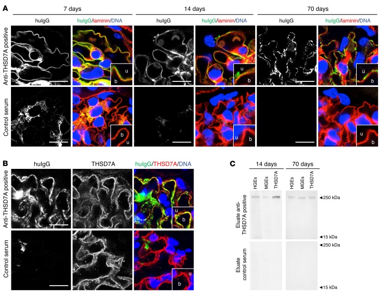
Figure 3. Anti-THSD7A antibodies induce the histological features of MN in mice.
(A) Immunofluorescence staining (paraffin sections) for huIgG and laminin in mice injected with anti-THSD7A antibody–containing or control serum at different time points. b, blood side of GBM; u, urinary side of GBM. Scale bars: 10 μm. Enlargements are of boxed areas (original magnification, ×4) of the glomerular filtration barrier. (B) Immunofluorescence staining (frozen sections) for huIgG and THSD7A in mice injected with anti-THSD7A antibody–containing serum or control serum. Scale bars: 10 μm. (C) Immunoblots of HGEs, MGEs, and recombinant mouse THSD7A with eluates from frozen kidney sections from mice that either received anti-THSD7A antibody–containing serum or control serum.

