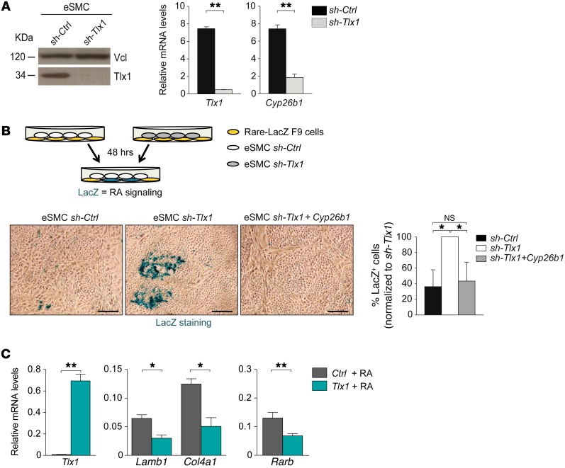
Figure 4. TLX1 controls RA activity.
(A) Western blot analysis of TLX1 protein in eSMCs upon shRNA silencing of Tlx1 (left). Anti-vinculin (Vcl) antibody is used as a loading control. Expression of Tlx1 and Cyp26b1 in sh-Ctrl and sh-Tlx1 eSMCs (right). Data are representative of 3 independent experiments. (B) Scheme of the coculture experiments with RARE-LacZ F9 reporter cells and sh-Ctrl or sh-Tlx1 eSMCs. Bright-field images of LacZ staining of F9-RARE-LacZ reporter cells cocultured for 48 hours with sh-Ctrl eSMCs (control), sh-Tlx1 eSMCs (silenced), or sh-Tlx1 eSMCs re-expressing Cyp26b1. Scale bars: 50 μm. Differences were measured by counting of the number of LacZ+ cells (in blue) over total cells. Data are representative of 1 of 3 independent experiments. (C) RARE-F9 cells transiently expressing Tlx1 or control vector were treated with RA and expression of indicated genes analyzed by qPCR 30 hours later. Data are representative of 1 of 3 independent experiments. (A–C)The means of triplicates ± SD are shown, *P < 0.05 (B and C), **P < 0.01 (A and C) (2-tailed Student’s t test).

