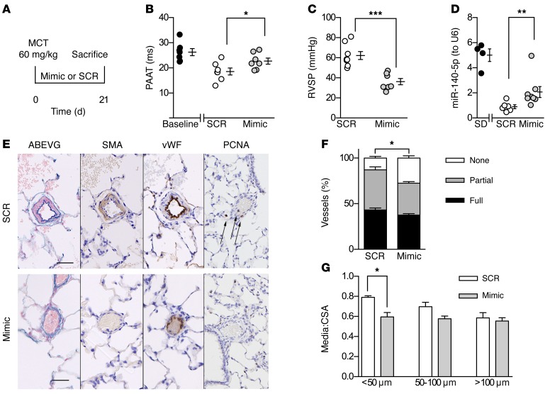
Figure 3. miR-140-5p prevents the development of PAH in the MCT rat model.
(A) Experimental time line. (B) PAAT at days 0 and 21. (C) RVSP at day 21. (D) qPCR showing whole lung miR-140-5p levels at day 21. (E) Representative photomicrographs of lung sections from SCR and miR-140-5p mimic–treated animals at day 21. Sections stained with Alcian blue EVG, α–smooth muscle actin (SMA), vWF, and PCNA (photomicrographs representative of n = 7–8 per group). Original magnification, ×200. Scale bars: 50 μm. (F and G) Pulmonary vascular remodeling by percentage muscularized vessels (F) and medial wall thickness as a ratio of total vessel size (media/CSA) (G) (B–G: n = 7–8 per group, *P < 0.05; **P < 0.01; ***P < 0.001, 2-tailed Mann-Whitney U test, mean ± SEM).

