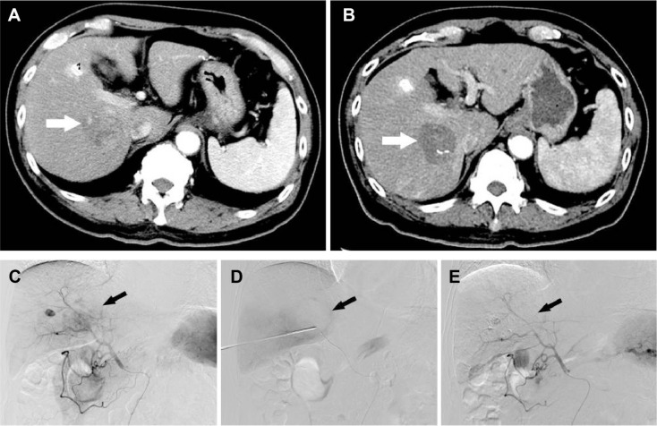
Figure 1.
A 75-year-old man who presented with CRLM treated with MWA combined with synchronous TACE.
Notes: (A) Enhanced CT scan of liver metastases in the right lobe of the liver, with a diameter of 4.2 cm ×3.5 cm (white arrow). (B) Three months after treatment, the enhanced CT scan showed a complete nonenhanced area of the tumor, indicating complete necrosis of the tumor lesion (white arrow). (C) DSA before ultrasound-guided percutaneous MWA combined with synchronous TACE clearly demonstrated the hypervascularity of the lesion (black arrow). (D) After the MWA, the lesion was almost eliminated (black arrow). (E) DSA showed complete devascularization after TACE (black arrow).
Abbreviations: CRLM, colorectal liver metastases; CT, computed tomography; DSA, digital subtraction angiography; MWA, microwave ablation; TACE, transcatheter arterial chemoembolization.

