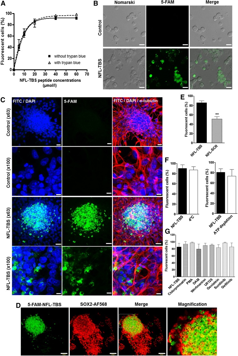
Figure 1.
Uptake of the NFL-TBS.40-63 peptide in neural stem cells from newborn rats. (A): The percentage of neural stem cells (NSCs) incorporating 5-FAM-labeled NFL-TBS.40-63 peptide, with (dark curve) and without (dotted curve) 0.4% trypan blue, was analyzed using the fluorescence-activated cell sorting technique. (B): Confocal microscopy of NSCs incubated with or without 20 µmol/l 5-FAM-labeled NFL-TBS.40-63 peptide (green). Scale bars = 10 µm. (C): Confocal microscopy of neurospheres incubated without (control) or with 20 µmol/l 5-FAM-labeled NFL-TBS.40-63 peptide (green) immunostained with anti-α-tubulin (red) to reveal the microtubule network. The nuclei were stained with DAPI (blue). Scale bars = 20 µm (original magnification ×63) and 5 µm (original magnification ×100). (D): Confocal microscopy of neurospheres incubated with 20 µmol/l 5-FAM-labeled NFL-TBS.40-63 peptide (green) immunostained with an anti-SOX2 antibody (red) to reveal stem cells. The nuclei were stained with DAPI (blue). Scale bars = 50 µm. (E): Percentages of NSCs that incorporate 5-FAM-labeled NFL-TBS.40-63 or scrambled peptides (20 µmol/l). Percentages of NSCs that incorporate 5-FAM-labeled NFL-TBS.40-63 peptide (20 µmol/l), after pretreatment in an ATP-depleted buffer or at 4°C (F) or with different endocytosis and signaling pathway inhibitors (G). Data are presented as mean ± SEM. ∗∗, p < .01. Abbreviations: 5-FAM, 5-carboxyfluorescein; DAM, 5-(N,N-dimethyl) amiloride hydrochloride; DAPI, 4′,6-diamidino-2-phenylindole; FITC, fluorescein isothiocyanate; PMA, phorbol 12-myristate 13-acetate.

