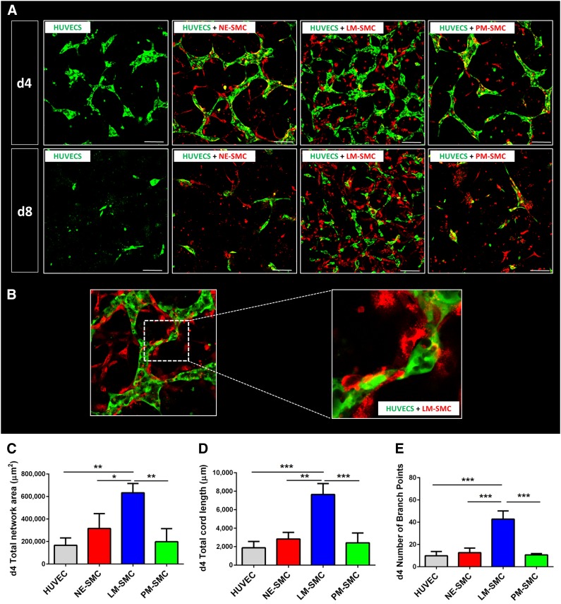
Figure 2.
LM-derived SMCs best support HUVEC network formation in a three-dimensional coculture model. (A): Confocal images of HUVEC (green) cocultures with the three respective SMC types (red) on days 4 and 8 of the coculture protocol. (B): SMCs provide physical support to developing networks, wrapping around the endothelial network. (C): Quantification of total network area. (D): Quantification of total cord length. (E): Quantification of number of branch points. Quantitative data are shown for day 4 of the coculture (∗, p < .05; ∗∗, p < .01; ∗∗∗, p < .001; n = 3 independent biological triplicates; scale bars = 100 μm). Abbreviations: d, day; HUVEC, human umbilical vein endothelial cell; LM, lateral mesoderm; NE, neuroectoderm; PM, paraxial mesoderm; SMC, smooth muscle cell.

