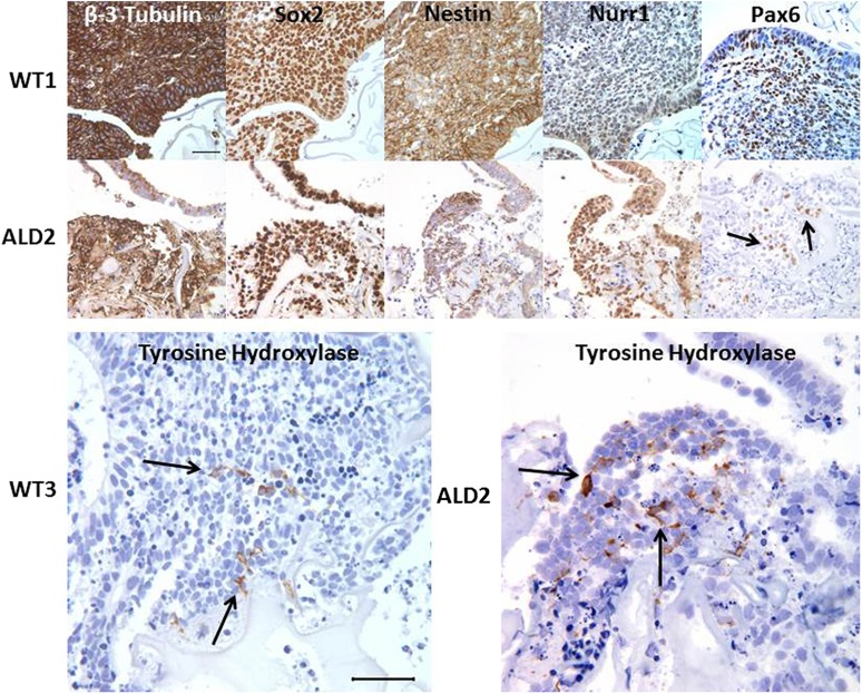
Figure 2.
Representative immunohistochemical staining panels for neural markers on day 14 cerebral organoids (cOrgs) derived from control (WT1 and WT3) or cerebral childhood adrenoleukodystrophy patient (ALD2) human induced pluripotent stem cells (hiPSCs). The majority of cells within all cOrgs derived from either control or adrenoleukodystrophy (ALD)-patient hiPSCs stained for β-3 tubulin, Sox2, and nestin. More localized expression of Nurr1 and Pax6 was seen in most cOrgs. Tyrosine hydroxylase positive cells, which often exhibited neuronal-like processes, were found in loose groupings in many of the cOrgs. Scale bars = 50 µm. Specific immunohistochemical stains (brown stain) with hematoxylin (blue) nuclear stain. Abbreviation: Nurr1, nuclear receptor related 1.

