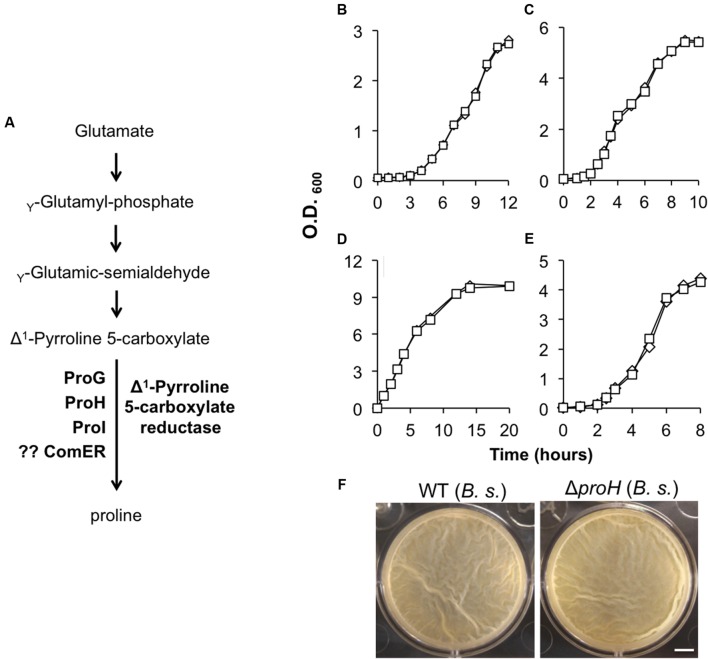
FIGURE 3.
(A) A schematic drawing of the proposed pathway for proline biosynthesis in B. subtilis. ProG, ProH, and ProI resemble the Δ1-pyrroline-5-carboxylate reductase and were shown to be involved in the biosynthesis of proline in B. subtilis (Belitsky et al., 2001). No evidence shows that ComER is also involved in the last step of proline biosynthesis in B. subtilis. (B,C) Growth of the WT (diamonds) and the comER mutant (squares) of B. cereus (B) and of B. subtilis (C) in MSgg. Assays were repeated multiple times and representative results were shown here. (D–E) Growth of the WT (diamonds) and the comER mutant (squares) of B. cereus (D) and of B. subtilis (E) in LBGM. Assays were repeated multiple times and representative results were shown here. (F) Pellicle biofilm formation by the WT (3610) and the ΔproH deletion mutant (B268) of B. subtilis in LBGM. Pictures were taken after 24 h of incubation at 30°C. The scale bar represents 5 mm in length.

