FIGURE 8.
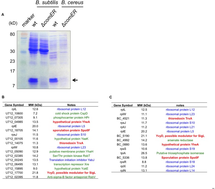
Candidate proteins that are differentially accumulated in the WT cells and the ΔcomER mutants of B. subtilis and B. cereus. (A) SDS-PAGE of the total protein lysates prepared from the WT strains and the ΔcomER mutants of B. subtilis and B. cereus. Cells were grown under shaking conditions in LBGM to early stationary phase (OD600 = 2). Major protein bands abundant in the lanes corresponding to the total lysates from the WT strains of B. subtilis and B. cereus, but largely absent in those from the comER mutants are indicated by the arrow. The size of the indicated proteins is estimated to be around 10–15 kD. (B–C) A list of the candidate proteins from samples of B. subtilis 3610 (B) and from B. cereus AR156 (C) based on MS analysis. Candidate proteins in blue represent ribosome or ribosome-associated proteins. Candidate proteins in green represent proteins that are present in both the WT samples and samples from the comER mutant (at lower levels). Candidate proteins (TrxA, YvyD, YheA, and Spo0F) in bold red are uniquely and also highly (relative counts above 10) present in the WT samples from both B. subtilis and B. cereus, but not in the samples from the comER mutants (Supplementary Tables S3–S6). Gene symbols were adopted from the NCBI database.

