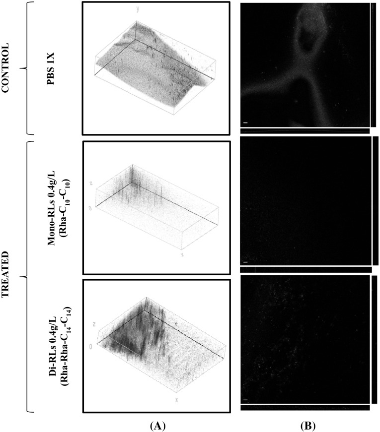
Fig. 4.
Confocal microscopy micrographs. a Three-dimensional (left panels) and b orthogonal reconstructions (right panels) of the biofilm formed by Bacillus subtilis BBK066. The pictures refer to the various experimental conditions as indicated on the left. The fluorescence is associated with live (green) and dead (red) cells, respectively. Scale bars represent 30 μm as indicated in micrographs (Color figure online)

