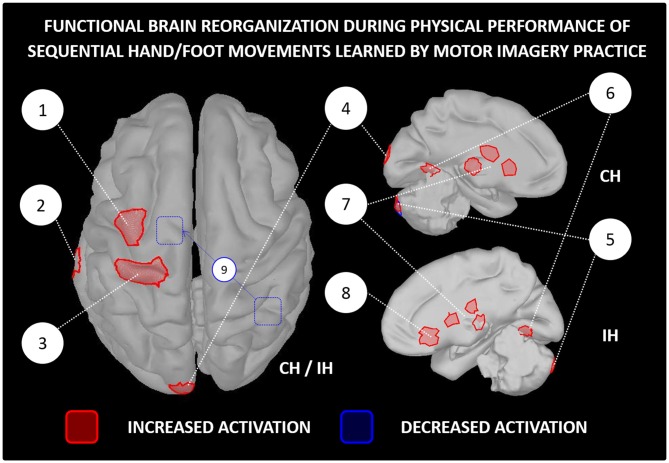
Figure 3.
Functional reorganization of the brain networks controlling the physical performance of a motor task learnt by MIP only. The figure is based on functional brain imaging experiments which performed source reconstruction analyses. Only paradigms involving sequential hand/foot movements met such inclusion criteria (e.g., Jackson et al., 2003; Lacourse et al., 2004; Nyberg et al., 2006; Zhang et al., 2011, 2012). Functional brain imaging experiments assessing neuroplasticity following MIP by examining brain activations during MI were not included (e.g., Sauvage et al., 2015). 1-Premotor cortex, 2-Middle temporal gyrus, 3-Primary motor cortex, 4-Occipital cortex, 5-Cerebellum, 6-Fusiform gyrus, 7-Thalamus and basal ganglia (caudate nucleus and putamen), 8-Orbitofrontal cortex, 9-Decreased functional connectivity between the right inferior parietal lobe and the supplementary motor area after MIP. MIP, Motor imagery practice; CH, Contralateral hemisphere; IH, Ipsilateral hemisphere.

