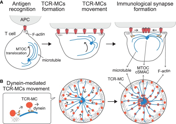
Figure 2.
Translocation of TCR–microclusters through cytoskeletal regulation. (A,B) depict the x–y axis and the z axis, respectively. Upon Ag recognition, T cells generate TCR–MCs (red) all over the interface. During this time, the MTOC (blue dot) is quickly translocated to the vicinity to the plasma membrane and finally to the TCR engagement site. Initially, TCR–MCs generated in the peripheral area move toward the center, coincident with actin retrograde flow (mesh structure). Thereafter, TCR–MCs are translocated to the cSMAC along the microtubules (blue line), which are translocated together with the MTOC into close proximity to the membrane in a dynein-mediated manner. The TCR/CD3 complex associates with the dynein–dynactin complex upon TCR stimulation, and then further assembles with microtubules. The dynein-mediated translocation of TCR–MCs regulates T cell activation because blockade of microtubules or dynein function prevents cSMAC formation and enhances T cell activation.

