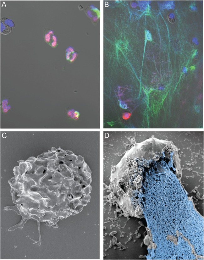
Figure 1.
NETs form during osmotic lysis of human neutrophils. (A) Immunofluorescence staining of freshly isolated human PMNs (histone 2A; red), MPO (green), and DNA (DAPI; blue). (B) NETs formed following electropermeabilization (pulse of 800 V at 25 mF). Brightness and contrast of the images in (A,B) were adjusted in Adobe Photoshop CC2014 (Adobe Systems Inc., San Jose, CA, USA). (C) Scanning electron micrograph of a control neutrophil that was not electropermeabilized, and (D) NET-forming human neutrophil following electropermeabilization (pulse of 600 V at 10 mF). Studies with human neutrophils were performed according to a protocol approved by the Institutional Review Board for Human Subjects, US NIAID/NIH, as described elsewhere (87). All subjects gave written informed consent prior to participation in the study and in accordance with the Declaration of Helsinki. The image in (A) was originally published in Ref. (87). Copyright © (2013) The American Association of Immunologists, Inc.

