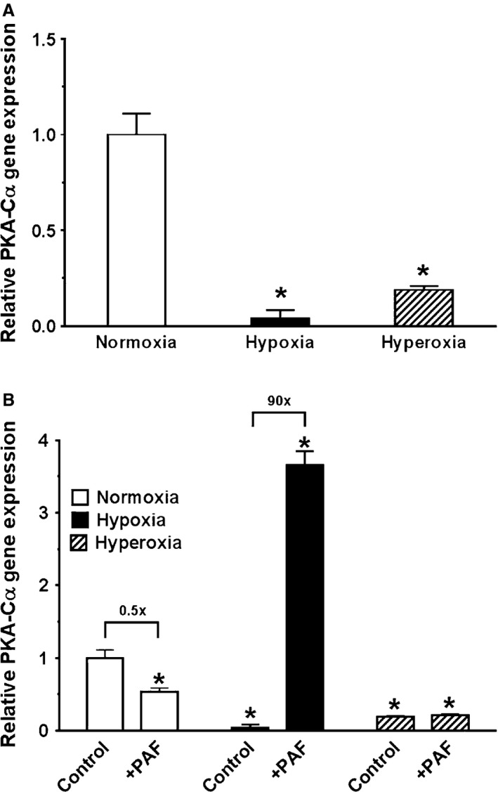
Figure 8.

A, Oxygen tension effect on PKA‐Cα gene expression, studied by qRT‐PCR in unstimulated newborn pulmonary artery smooth muscle cells (PASMC). RNA from newborn PASMC was studied by quantitative RT‐PCR (qRT‐PCR) studies to examine PKA‐Cα gene expression by cells cultured in normoxia, hypoxia, and hyperoxia under baseline conditions. Gene expression was normalized to expression of GAPDH internal standard. Data are mean ± SEM, n = 3 different cell preparations. Both exposure to hypoxia and hyperoxia decreased PKA‐Cα gene expression significantly (hypoxia by 96% and hyperoxia by 81%, respectively). The statistics are: *P < 0.05, different from normoxia. B, Oxygen tension effect on PKA‐Cα gene expression, studied by qRT‐PCR in controls and treatments with platelet‐activating factor (PAF). The results of PKA‐Cα gene expression in unstimulated controls from Figure 6a are presented here in comparison with newborn PASMC that were concurrently stimulated with exogenous 10 nmol/L PAF. All values were calculated relative to untreated normoxia‐exposed controls. Ratios of gene expression in untreated to treated cells were calculated within each oxygen tension and presented in fold difference above brackets. Gene expression was normalized to expression of GAPDH internal standard. Data are mean ± SEM, n = 3 different cell preparations. The greatest PKA‐Cα gene expression yielded from PAF‐stimulated cells in hypoxia (3.6 times greater than unstimulated controls in normoxia). PKA‐Cα gene expression was significantly decreased by hyperoxia exposure, regardless of treatment with PAF. Stimulation with exogenous PAF decreased PKA‐Cα gene expression in normoxia by 50%. The statistics are: *P < 0.05, different from normoxia‐exposed untreated controls.
